Centre for Virus research, Therapeutics and vaccines
Translational Health Science and Technology Institute, Faridabad-121001, India


Research
Host-pathogen interactions in HEV and SARS-CoV-2 infected cells
Host-pathogen interactions determine the survival or extinction of the host and the pathogen. This eternal conflict between the host and the pathogen is a major driving force in their evolution. Therefore, a thorough understanding of the molecular details of the host-pathogen conflicts are crucial for decoding their survival skills and developing specific therapeutics against the pathogen.

HEV-host protein interaction network
Toward understanding the molecular details of HEV life cycle, we screened human liver and fetal brain cDNA libraries to identify the host interaction partners of proteins encoded by genotype 1 HEV and constructed the virus-host PPI network. Analysis of the network indicated a role of HEV proteins in modulating multiple host biological processes such as stress and immune responses, the ubiquitin-proteasome system, energy and iron metabolism, and protein translation. Further investigations revealed the presence of multiple host translation regulatory factors in the viral translation/ replication complex. Depletion of host translation factors such as eIF4A2, eIF3A, and RACK1 significantly reduced the viral replication, whereas eIF2AK4 depletion had no effect. These findings highlight the ingenuity of the pathogen in manipulating the host machinery to its own benefit, a clear understanding of which is essential for the identification of strategic targets and development of specific antivirals against HEV.

IRES-element mediated translation of the ORF4 protein promotes g1-HEV replication
Multiple processes exist in a cell to ensure continuous production of essential proteins either through cap-dependent or cap-independent translation processes. Viruses depend on the host translation machinery for viral protein synthesis. Therefore, viruses have evolved clever strategies to utilize the host translation machinery. Our studies show that genotype 1-Hepatitis E virus (g1-HEV) utilizes both cap-dependent and cap-independent translation machineries for its translation and proliferation. Cap-independent translation in g1-HEV is driven by an eighty seven nucleotide-long RNA element which acts as a noncanonical, internal ribosome entry site like (IRESl) element. HEV IRESl associates with several host ribosomal proteins for its function. Our findings demonstrate indispensable roles of ribosomal protein RPL5 and DHX9 (RNA helicase A) in mediating HEV IRESl activity. HEV IRESl drives the expression of viral protein ORF4 in an endoplasmic reticulum stress-inducible manner. Importantly, ORF4 expression and stimulatory effect of ER stress inducers on viral replication is specific to g1-HEV. ORF4 protein sequence is mostly conserved among g1-HEV isolates and ORF4 specific antibodies are detected in g1-HEV patient serum. ORF4 interacts with multiple viral and host proteins and facilitates the assembly of a protein complex consisting of viral helicase, RNA dependent RNA polymerase (RdRp), X, host eEF1α1 (eukaryotic elongation factor 1 isoform-1) and tubulinβ. In association with eEF1α1, ORF4 stimulates viral RdRp activity.

Proviral Function of HEV Protease via eIF2AK4 Inhibition
Data obtained from HEV protein interactome study reveals the interaction between viral protease (PCP) and cellular eukaryotic initiation factor 2 alpha kinase 4 (eIF2AK4). Since eIF2AK4 is a sensor of the cellular integrated stress response machinery, we evaluated the significance of the interaction between PCP and eIF2AK4. PCP was found to competitively associate with and interfere with self-association of the eIF2AK4, thereby inhibiting its kinase activity. Lack of eIF2AK4 activity prevents phosphorylation-mediated inactivation of the cellular eIF2α, which is essential for initiation of cap-dependent translation. Thus, PCP behaves as a proviral factor, promoting uninterrupted synthesis of viral proteins in infected cells, which is crucial for survival and proliferation of the virus.

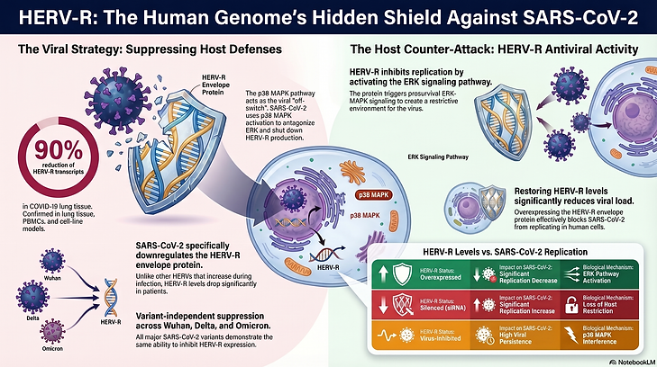
Deciphering the antiviral role of HERV-R -envelope against SARS-CoV-2
Human endogenous retroviruses (HERVs) represent retroviral elements that were integrated into the ancestral human genome. HERVs are important in embryonic development as well as in the manifestation of diseases, including cancer, inflammation, and viral infections. We observed down regulation of HERV-R envelope in cell-based models and PBMCs of COVID-19 patients. Overexpression of HERV-R inhibits SARS-CoV-2 replication, suggesting its antiviral activity. Further analyses demonstrate the role of the extracellular signal-regulated kinase (ERK) in regulating HERV-R antiviral activity. Lastly, we demonstrate that the crosstalk between ERK and p38 MAPK controls the synthesis of the HERV-R envelope protein, which in turn modulates SARSCoV-2 replication. These findings suggest the role of the HERV-R envelope as a prosurvival host factor against SARS-CoV-2 and illustrate a possible advantage of integration and evolutionary maintenance of retroviral elements in the human genome.
SARS-CoV-2 5'-3'-UTR-RNA-protein interactome
Replication of a positive-strand RNA virus involves an RNA-protein complex consisting of viral genomic RNA, host RNA(s), virus-encoded proteins, and host proteins. Dissecting out individual components of the replication complex will help decode the mechanism of viral replication. 5'- and 3'-UTRs in positive-strand RNA viruses play essential regulatory roles in virus replication. Using an RNA-protein interaction detection (RaPID) assay coupled to liquid chromatography with tandem mass spectrometry, we identified host interaction partners of SARS-CoV-2 5'- and 3'-UTRs and generated an RNA-protein interaction network. By combining these data with the previously known protein-protein interaction data proposed to be involved in virus replication, we generated the RNA-proteinprotein interaction (RPPI) network, likely to be essential for controlling SARS-CoV-2 replication. Notably, bioinformatics analysis of the RPPI network revealed the enrichment of factors involved in translation initiation and RNA metabolism. Lysosome-associated membrane protein-2a (Lamp2a), the receptor for chaperone-mediated autophagy, is one of the host proteins that interact with the 5'-UTR. Further studies showed that the Lamp2 level is upregulated in SARS-CoV-2-infected cells and that the absence of the Lamp2a isoform enhanced the viral RNA level whereas its overexpression significantly reduced the viral RNA level. Lamp2a and viral RNA colocalize in the infected cells, and there is an increased autophagic flux in infected cells, although there is no change in the formation of autophagolysosomes. In summary, our study provides a useful resource of SARS-CoV-2 5'- and 3'-UTR binding proteins and reveals the antiviral role of Lamp2a protein during SARS-CoV-2 infection.

Towards New Antivirals

Drug repurposing study identifies potential anti-HEV activity of Etravirine
Computational biology tools promise to revolutionise the antiviral drug discovery. We analyzed the transcriptome data of HEV infected primary human hepatocyte (PHH)-cells through connectivity map database and applied control theory on functional network to identify antiviral targets against HEV. PKCβ, PKB/AKT and CK1ε were identified as potential antiviral targets against HEV. Antiviral function of PKB/AKT and CK1ε was confirmed using cell-based models of genotype 1 (g1)- and g3-HEV infection. Further experiments demonstrated the antiviral activity of Etravirine against HEV, mediated via its ability to inhibit the CK1ε activity. Etravirine is an FDA approved non-nucleoside reverse transcriptase inhibitor, used for the treatment of Acquired immunodeficiency syndrome (AIDS) patients. This study reveals the potential of repurposing Etravirine for treatment of HEV patients and illustrate the importance of computational biology in antiviral drug discovery.

Antiviral activity of Zinc against HEV
Zinc is an essential micronutrient that plays crucial roles in multiple cellular processes. Zinc also acts as a broad spectrum antimicrobial agent. While searching for antivirals against HEV, we observed potent antiviral activity of Zinc salts against HEV. Zinc oxide (ZnO) and its nanostructures have attracted marked interest due to their unique characteristics. Weevaluated antiviral activity of ZnO nanoparticles [ZnO(NP)] and tetrapod-shaped ZnO nanoparticles [ZnO(TP)] against HEV. Both ZnO(NP) and ZnO(TP) displayed potent antiviral activity against HEV, the latter being more effective. Measurement of cell viability and intracellular Reactive Oxygen Species levels revealed that both ZnO(NP) and ZnO(TP) are noncytotoxic to the cells even at significantly higher dose, compared to a conventional Zinc salt (ZnSO4).Interestingly, ZnO(TP) nanoparticles are also effective against HCV. Our study paves the way for evaluation of potential therapeutic benefit of ZnO(TP) against HEV and HCV.

Viral vaccines
Viral hepatitis is a major public health concern and WHO aims at eliminating it by 2030. Hepatitis E virus (HEV) is a major cause of acute viral hepatitis (AVH) and community level outbreaks. It may also cause chronic and fulminant hepatitis. The disease worsens during pregnancy, with a 20-25% mortality rate. Recent reports suggest chronic HEV infection in ~60% of liver transplantation patients. My laboratory is engaged in developing a recombinant virus-like particle (VLP)-based vaccine against the HEV.
We have also established an mRNA vaccine development platform to address the requirement of vaccines against pathogens of human medical importance. The platform utilizes the sequence information of the potential vaccine antigens of any pathogen to produce a series of lipid nanoparticle (LNP) encapsulated-mRNA vaccine formulations, which are evaluated in cell line-based and small animal model systems to identify the best vaccine antigen. Proof of function of the mRNA vaccine development platform was shown in our laboratory by producing and evaluating vaccine candidates against different variants of SARS-CoV-2. Our experimental conditions produced circular mRNA (cRNA)-LNP vaccine formulations encoding RBD of Wuhan, Delta and Omicron (BA.4/5) variants of SARS-CoV-2, with an average particle size of 136nm, PDI of 0.05 and 96% cRNA encapsulation efficiency. The average end point IgG (anti-RBD) and Neutralization (by PRNT assay) titer of the different RBD variants was in the range of 10000-100000 and 1000-10000, respectively. Moreover, single variant vaccine formulations as well as a bivalent cRNA vaccine formulation [encoding RBD of Delta and Omicron (BA.4/5) variants] protected against corresponding SARS-CoV-2 variant challenges in mouse model of COVID-19, thus confirming the functional efficacy of the vaccine formulations.
